Peptides and Proteins
A concise revision guide to peptides and proteins: the formation of the peptide (amide) link by condensation between amino acids, drawing dipeptides and tripeptides, the hydrolysis of proteins to their amino acids and their separation by chromatography, and the primary, secondary and tertiary structure of proteins and how enzymes work.
Forming a Peptide Link
Choose a step to watch two amino acids lose water, form a peptide link, grow into a chain and split apart again on hydrolysis.
© Dr. Mohammed Al-Fatah – onlinelearningsystem.net
Before You Start
A peptide is a chain of amino acids joined by amide links. Check these ideas first.
- The general formula RCH(NH₂)COOH, the R groups of glycine, alanine and serine, and the zwitterion: Amino Acids and Zwitterions.
- The amide group –CONH– and how an acyl chloride and an amine form it with loss of a small molecule: Amines as Nucleophiles.
- A condensation reaction joins two molecules and releases a small molecule, here water.
- A condensation polymer forms when every monomer has two reactive groups: Condensation Polymers: Polyamides and Polyesters.
GCSE Recap: Polymers and Their Monomers
Three quick questions on the GCSE ideas this page builds on.
The Peptide Link
The –COOH group of one amino acid and the –NH₂ group of another join with the loss of water. The new –CONH– group is an amide link, called the peptide link in peptides and proteins.
- Glycine + alanine → a dipeptide + H₂O: two amino acids, one peptide link.
- Two different dipeptides are possible. In Gly-Ala the glycine provides the C=O and the alanine the N–H; in Ala-Gly it is the other way round.
- H₂NCH₂COOH + H₂NCH(CH₃)COOH → H₂NCH₂CONHCH(CH₃)COOH + H₂O (Gly-Ala).
- Every dipeptide still has a free –NH₂ at one end and a free –COOH at the other, so the chain can keep growing.
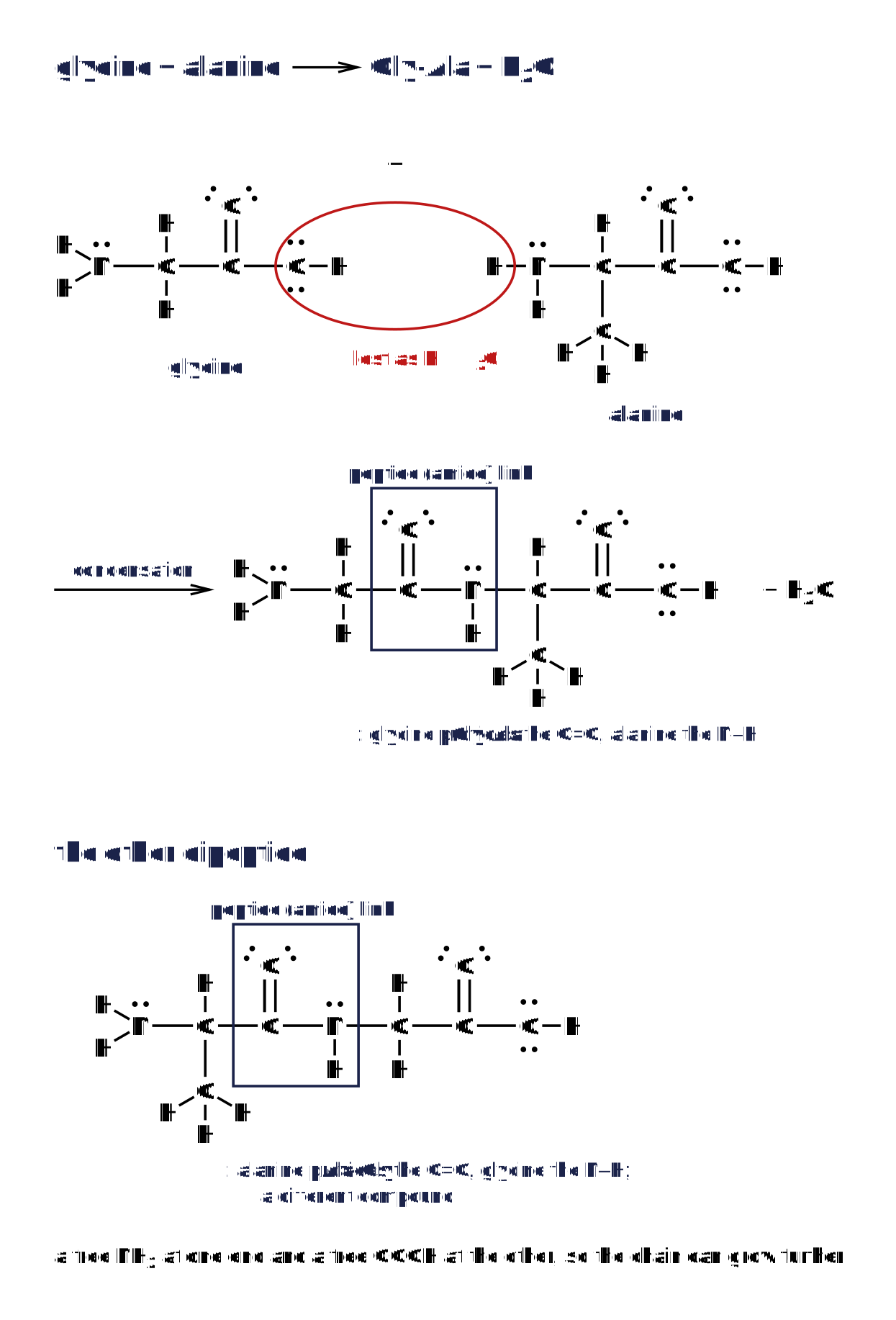
Glycine and alanine condensing to the Gly-Ala dipeptide with the peptide link boxed, and the isomeric Ala-Gly dipeptide underneath.
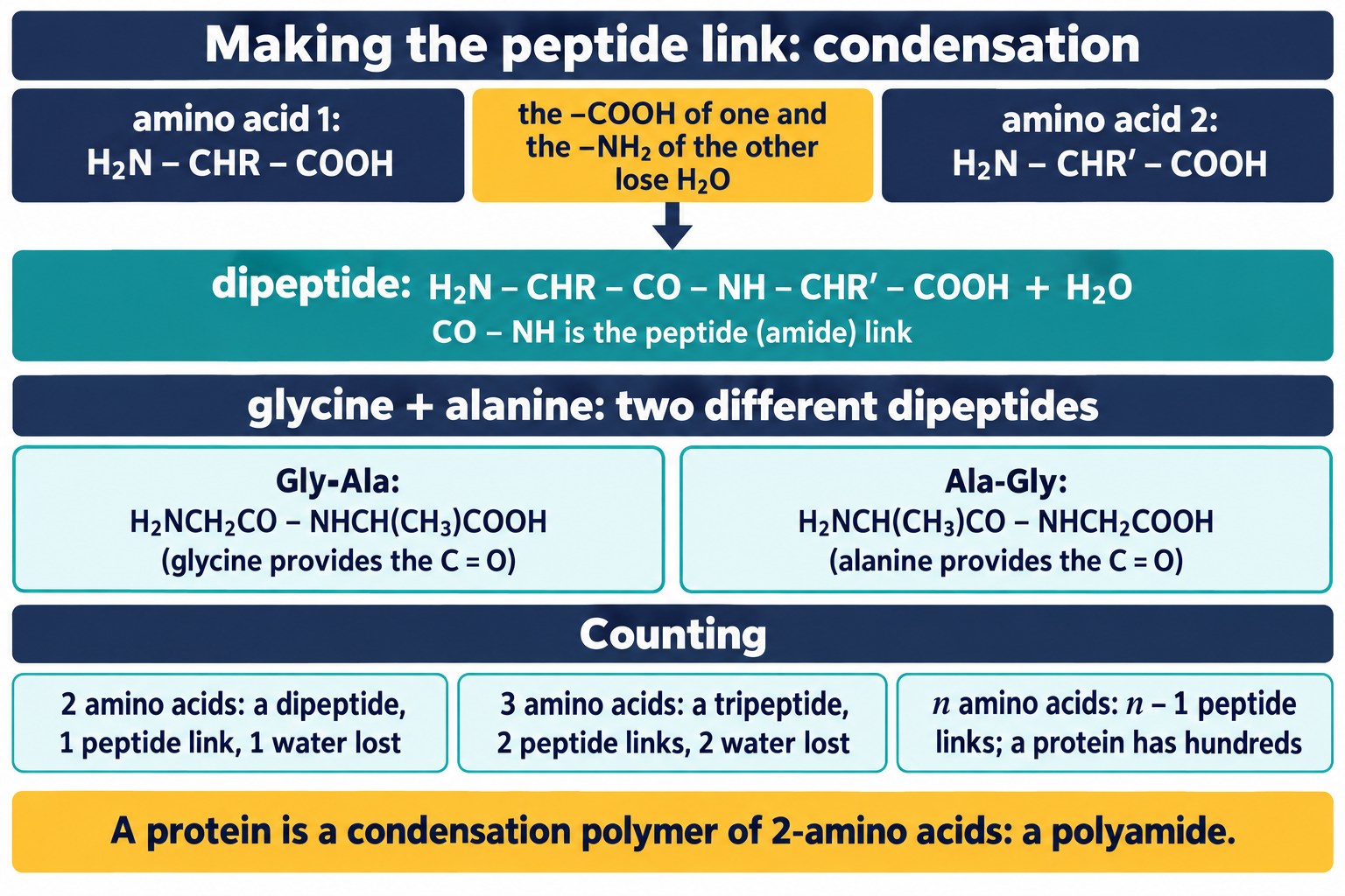
Making the peptide link: the acid of one amino acid and the amine of another lose water, the two dipeptides from glycine and alanine, and how to count the links in a chain.
Exam focus: When two different amino acids are given, say that two dipeptides can form and draw both. The order matters: Gly-Ala and Ala-Gly are different compounds.
Tripeptides and Longer Chains
Three amino acids give a tripeptide with two peptide links; n amino acids give n − 1 links and lose n − 1 water molecules.
Worked example: drawing a tripeptide
Step 1. Draw glycine, alanine and serine in order, each with the –NH₂ on the left and the –COOH on the right: H₂NCH₂COOH, H₂NCH(CH₃)COOH, H₂NCH(CH₂OH)COOH.
Answer. Gly-Ala-Ser: H₂NCH₂CONHCH(CH₃)CONHCH(CH₂OH)COOH, two peptide links, two water molecules lost.
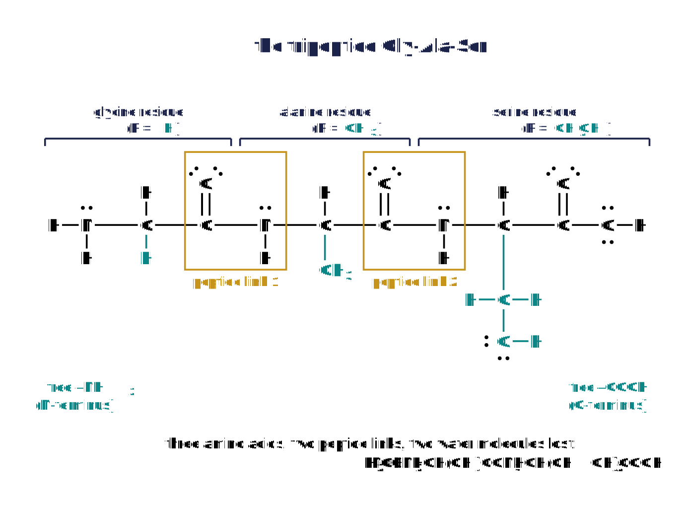
The Gly-Ala-Ser tripeptide drawn displayed with both peptide links boxed and the three residues bracketed and named.
Worked example: reading a peptide
Answer. The R groups are CH₂SH, H and CH₃: cysteine, glycine and alanine, in that order from the –NH₂ end.
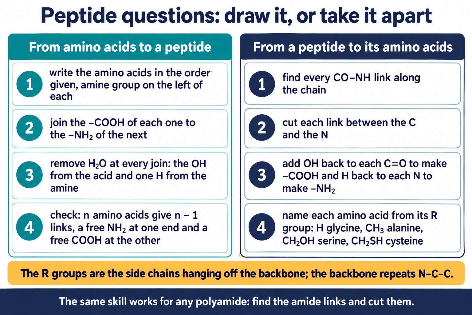
Peptide questions both ways: building a peptide from named amino acids, and taking a peptide apart to recover and name its amino acids.
Quick Check: Counting Sequences, Links and Mr
Type a whole number for each one.
Polypeptides and Proteins
A chain of many amino acids is a polypeptide; a protein is one or more long polypeptide chains. Proteins are condensation polymers of 2-amino acids.
- Each monomer has two reactive groups (–NH₂ and –COOH), so the chain grows by repeated condensation with loss of water at every link.
- A protein is therefore a polyamide, like nylon, with the repeating backbone –NH–CHR–CO–: Condensation Polymers: Polyamides and Polyesters.
- The sequence of amino acids along the chain, held by the peptide links, is the primary structure of the protein (below).
- About twenty different amino acids, in chains hundreds long, give an almost unlimited number of proteins.
Remember: Dipeptide: two amino acids, one link. Tripeptide: three, two links. Polypeptide and protein: many amino acids, a condensation polymer, a polyamide.
Check: Peptides
Draw the dipeptides from two given amino acids, count the peptide links in a chain, and identify the amino acids in a given peptide.
Breaking the Peptide Link: Hydrolysis
Heating a protein with acid or alkali breaks every peptide link by hydrolysis: water is added back across the C–N bond and the amino acids are released.
- Conditions: heat under reflux with 6 mol dm⁻³ hydrochloric acid for about 24 hours.
- In acid the amino acids are present as their ammonium salts, H₃N⁺CHRCOOH Cl⁻; neutralising gives the free amino acids.
- Alkaline hydrolysis with hot sodium hydroxide gives the carboxylate salts, H₂NCHRCOO⁻ Na⁺, instead.
- Gly-Ala + H₂O → glycine + alanine: one water molecule is added back for every link broken.
- The mixture of amino acids is then separated and identified by chromatography (below).
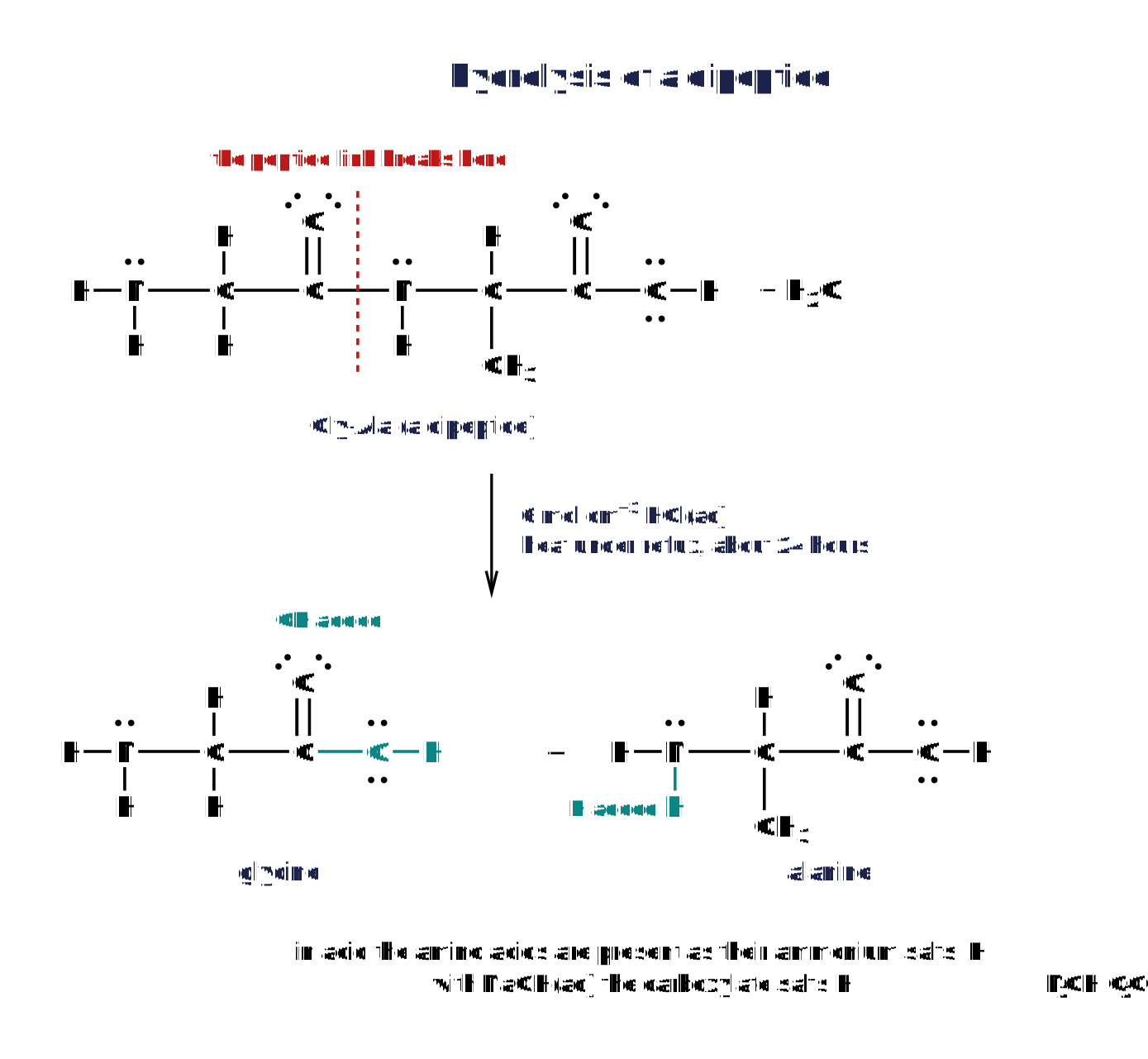
The Gly-Ala dipeptide hydrolysed under reflux with hydrochloric acid to glycine and alanine, with the bond that breaks marked.
Exam focus: Hydrolysis is the reverse of condensation: add H₂O across each CO–NH link, giving a –COOH and an –NH₂. State the acid (or alkali) and heat under reflux.
Quick Check: Hydrolysing a New Peptide
Pick the one accurate statement about each hydrolysis.
Separating the Amino Acids: Chromatography
The amino acids from a hydrolysed protein are separated by thin-layer chromatography (or paper chromatography) and identified by their Rf values: Chromatography and Rf Values.
- A spot of the mixture is placed on the baseline of a TLC plate, which stands in a solvent; the amino acids move up at different rates.
- Amino acids are colourless, so the dried plate is sprayed with ninhydrin, which turns the spots purple, or viewed under ultraviolet light.
- Rf = (distance moved by the spot) / (distance moved by the solvent front), both measured from the baseline.
- Each amino acid is identified by comparing its Rf with the values of known amino acids run in the same solvent.
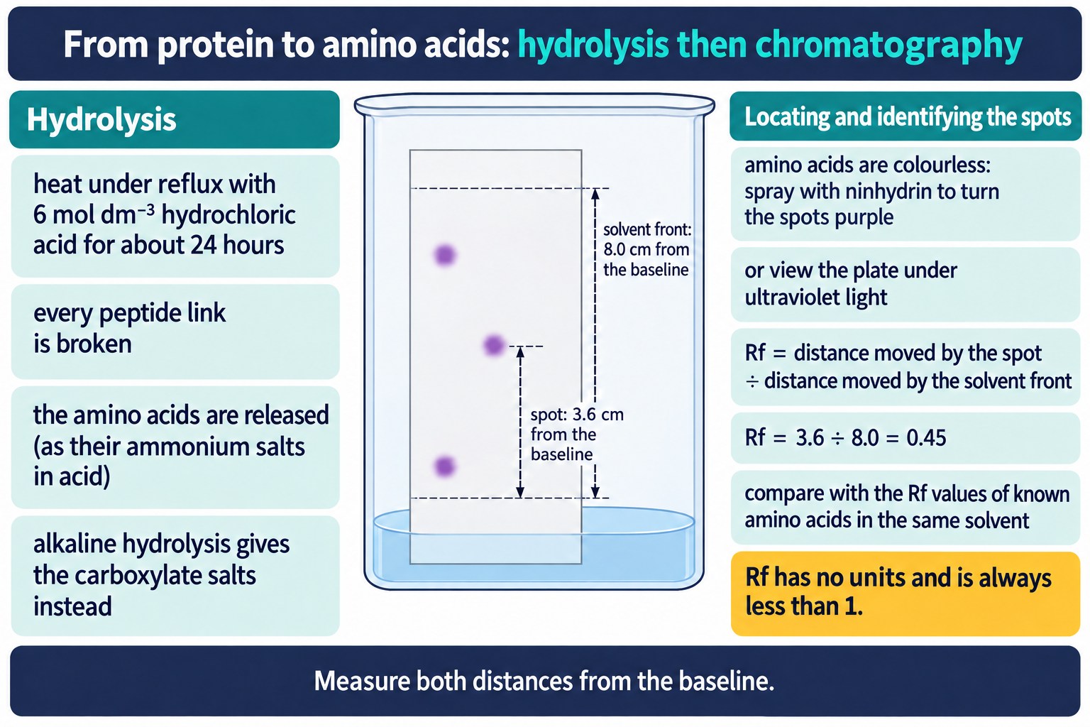
From protein to amino acids: reflux with 6 mol dm⁻³ hydrochloric acid, then a thin-layer chromatography plate with spots located by ninhydrin and an Rf calculation.
Worked example: an Rf value
Step 2. Rf = (3.6) / (8.0) = 0.45 (no units).
Remember: Rf is always between 0 and 1 and has no units. Measure to the centre of the spot, from the pencil baseline.
Protein Structure: Primary, Secondary, Tertiary
A protein has structure at three levels. Each level is held by a different kind of bonding.
| Level | What it is | Held by |
|---|---|---|
| primary structure | the sequence of amino acids along the chain | peptide links (covalent) |
| secondary structure | local shapes: the α-helix (a coil) and the β-pleated sheet (chains side by side) | hydrogen bonds between the C=O of one peptide link and the N–H of another |
| tertiary structure | the folding of the whole chain into its 3D shape | hydrogen bonds, ionic interactions, disulfide (S–S) bonds and van der Waals forces between side chains |
- Hydrogen bonds: the δ− oxygen of a C=O and the δ+ hydrogen of an N–H, in the backbone (secondary) or between side chains (tertiary).
- Ionic interactions: between an –NH₃⁺ side chain (lysine) and a –COO⁻ side chain (glutamic acid).
- Disulfide bonds: two cysteine residues are oxidised, their –SH groups joining as a covalent S–S bridge.
- Van der Waals forces: between non-polar side chains such as CH₃ and CH₂C₆H₅.
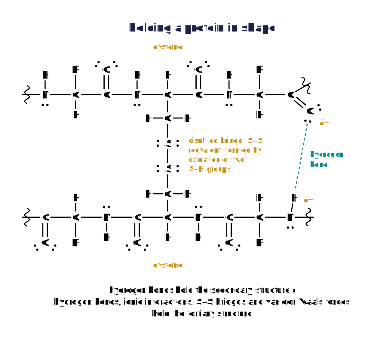
Two neighbouring chain fragments joined by a disulfide bridge between cysteine residues, with a hydrogen bond between a C=O and an N–H of two peptide links.
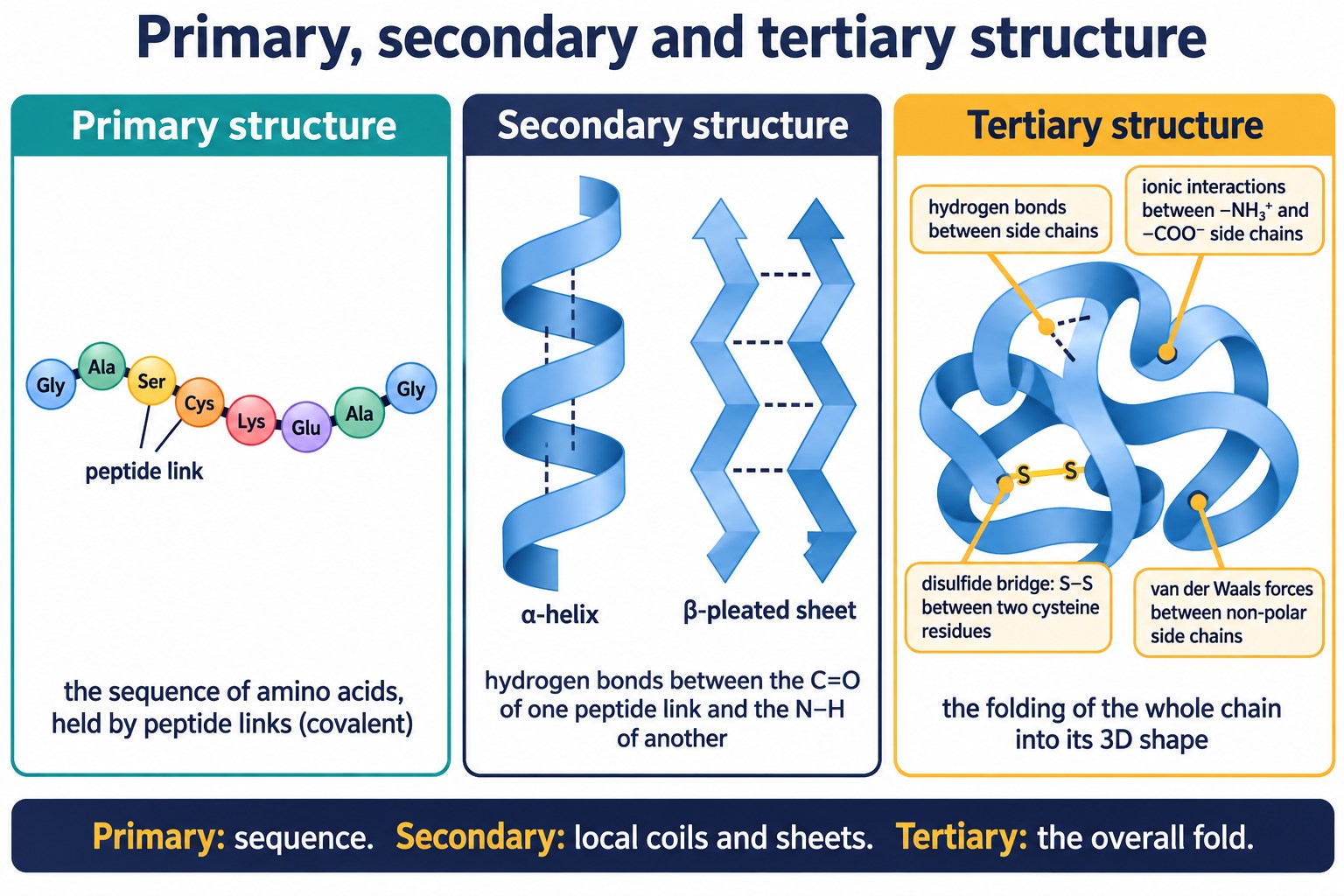
Primary sequence as a string of named beads, the α-helix and β-pleated sheet held by hydrogen bonds, and the folded tertiary structure with its four kinds of interaction.
Identifying the level in a diagram
- A list or string of amino acid names: primary structure.
- A coil or a set of parallel zigzag strands with hydrogen bonds between C=O and N–H: secondary structure.
- A whole folded chain with side-chain interactions marked: tertiary structure.
Exam focus: Name the interaction and the groups involved: “hydrogen bond between the C=O of one peptide link and the N–H of another” or “S–S bond between two cysteine residues”.
Quick Check: Naming the Interaction and Level
Identify the interaction or structural level in each new case.
Enzymes
Enzymes are proteins that act as catalysts. The reaction happens at the active site, a pocket in the tertiary structure whose shape and groups fit one substrate.
- The active site is stereospecific: it is itself chiral, so only one enantiomer of the substrate (or of a drug) has the right shape to bind.
- The other enantiomer does not fit and is not acted on, which is why one enantiomer of a drug can be active and the other inactive.
- A drug can act as an enzyme inhibitor: it binds in the active site and blocks it, so the substrate cannot bind and the reaction stops.
- Computers model the shape of the active site and predict which molecules will fit, helping chemists design inhibitor drugs before making them.
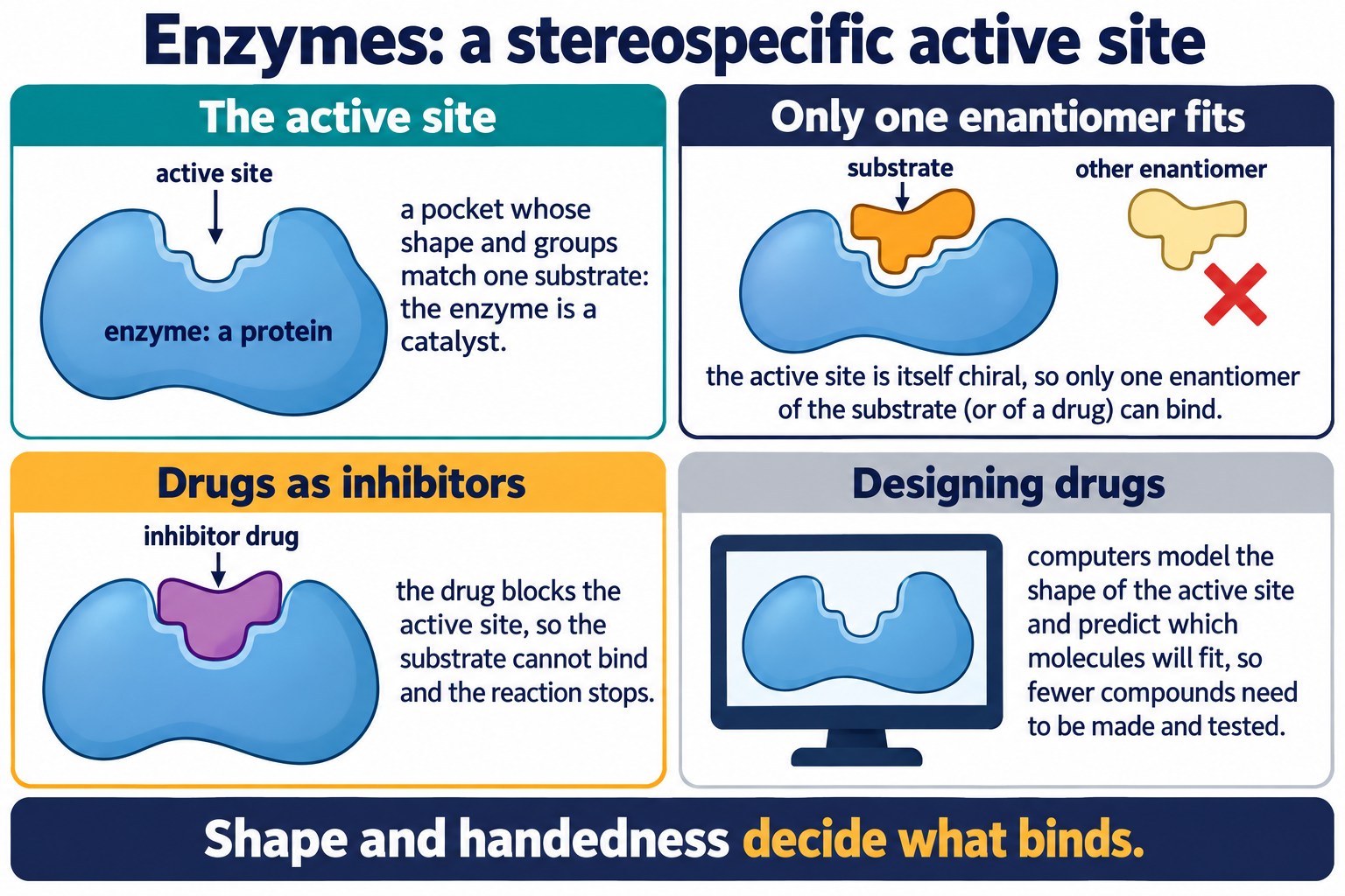
Lock and key in simple shapes: the active site, the substrate that fits and its enantiomer that does not, an inhibitor drug blocking the site, and computer-aided design.
Remember: Three words to use: catalyst, active site, stereospecific. Then: the inhibitor blocks the active site; the active site binds only one enantiomer.
Check: Hydrolysis, Chromatography and Proteins
Give the conditions for hydrolysing a protein, identify the amino acids formed, and recall what holds each level of protein structure together.
Common Exam Points
- The peptide link is an amide link, –CONH–, formed by condensation between the –COOH of one amino acid and the –NH₂ of another with loss of H₂O.
- Two different amino acids give two dipeptides (Gly-Ala and Ala-Gly); n amino acids give n − 1 peptide links.
- To draw a peptide, line up the amino acids and remove H₂O at every join; to recover them, cut each CO–NH link and add back OH and H.
- Proteins are condensation polymers of 2-amino acids: polyamides with the backbone –NH–CHR–CO–.
- Hydrolysis: heat under reflux with 6 mol dm⁻³ hydrochloric acid for about 24 hours; in acid the amino acids form as their ammonium salts.
- The amino acids are separated by thin-layer chromatography, located with ninhydrin (purple) or under UV light, and identified by Rf values (Rf = (spot distance) / (solvent front distance)).
- Primary: the sequence (peptide links). Secondary: α-helix and β-pleated sheet (hydrogen bonds between C=O and N–H). Tertiary: the overall fold (hydrogen bonds, ionic, S–S, van der Waals).
- Enzymes are protein catalysts with a stereospecific active site that binds one enantiomer; a drug can inhibit an enzyme by blocking the active site; computers help design such drugs.
FAQs
Use these quick answers to check the peptide link, the two dipeptides from a pair of amino acids, counting links, the hydrolysis of proteins, protein structure and enzymes.
What is a peptide link?
It is the amide link, –CONH–, formed when the COOH group of one amino acid condenses with the NH₂ group of another, losing a water molecule. The C=O comes from the acid group and the N–H from the amine group of the other.
Why can glycine and alanine form two different dipeptides?
It depends on which amino acid provides the C=O of the link. If the COOH of glycine reacts with the NH₂ of alanine, the dipeptide is Gly-Ala, H₂NCH₂CONHCH(CH₃)COOH.
If the COOH of alanine reacts with the NH₂ of glycine, it is Ala-Gly, a different compound.
Why are enzymes stereospecific?
An enzyme is a protein whose tertiary structure creates an active site of a particular shape. Only a substrate with the matching shape, and the correct enantiomer, can bind there. Drugs designed as inhibitors block the active site; computers model the site to design them.
How is a protein broken down into its amino acids?
Heat the protein under reflux with 6 mol dm⁻³ hydrochloric acid for about 24 hours. Water, catalysed by the acid, breaks every peptide link (hydrolysis) and the amino acids are released as their ammonium salts. Hot sodium hydroxide gives the carboxylate salts instead.
What is the difference between primary, secondary and tertiary structure?
Primary: the sequence of amino acids joined by peptide links. Secondary: the α-helix or β-pleated sheet, held by hydrogen bonds between C=O and N–H groups of peptide links.
Tertiary: the folding of the whole chain, held by hydrogen bonds, ionic interactions and disulfide bonds.
Copyright notice: This OLS revision content, including the explanations, layout, diagrams, tables and embedded learning structure, is authored for Online Learning System by Dr. Mohammed Al-Fatah. It may not be copied, reproduced, redistributed or adapted without written permission.
Keep this note — free
Save your progress across every AQA topic. A free account remembers which topics you have covered, saves your question scores, and syncs across your phone and laptop.
- Track every topic you have finished
- Keep your practice-question scores
- No payment, no card, free forever
Already registered? Log in