The Mass Spectrometer
A concise revision guide to how a mass spectrometer works, including vaporisation, ionisation by electron bombardment, acceleration, deflection, detection and the interpretation of m/z values.
Mass Spectrometer: Follow the Ions
Choose a step to follow the ions through the instrument: ionisation, acceleration, deflection, detection and changing the magnetic field.
© Dr. Mohammed Al-Fatah – onlinelearningsystem.net
Before You Start
This page builds on GCSE atomic structure: isotopes, ions and the forces between charges.
- Isotopes are atoms of the same element with the same number of protons but different numbers of neutrons.
- An atom that loses an electron becomes a positive ion.
- Opposite charges attract and like charges repel.
- The relative atomic mass of an element is a mean that allows for the abundance of each isotope.
GCSE Recap: Isotopes, Ions and Charges
Before you read on, check the GCSE ideas about isotopes, ions and charges that the instrument relies on.
What a Mass Spectrometer Does
A mass spectrometer separates positive ions according to their mass-to-charge ratio, written as m/z.
It is used to identify isotopes, compare their relative abundance, and provide data used to calculate relative atomic mass. The sample must be converted into particles that can move through the machine and be detected as ions.
The main sequence is: vaporisation, ionisation, acceleration, deflection and detection.
Key idea: The mass spectrometer does not directly weigh atoms. It forms positive ions, separates them by m/z, and records detector signals that show relative abundance.
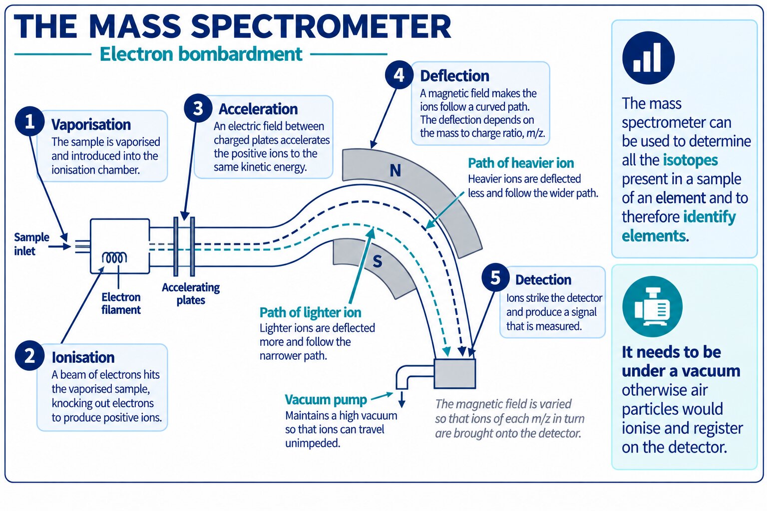
The detector records ions that pass through the selected path, allowing the computer to build a spectrum from their m/z values and abundances.
Why the Instrument Uses a Vacuum
The mass spectrometer operates at low pressure and under a vacuum.
This matters because air particles would otherwise collide with the ions, change their path, or become ionised themselves. If air particles reached the detector, they could produce extra signals and reduce the reliability of the spectrum.
Exam focus: A vacuum prevents collisions with air particles and prevents air particles from being ionised and detected.
Quick Check: What If Air Leaks In?
Explain what would go wrong if air leaked into the instrument.
Stage 1: Vaporisation and Ionisation
The sample must be in the gaseous state so that its particles can move through the mass spectrometer. If the sample is not already gaseous, it is first vaporised.
In electron bombardment, an electron gun fires high-energy electrons at the gaseous atoms or molecules. These electrons collide with the sample and remove an outer electron, forming a positive ion.
For an atom X, this can be represented as: X(g) + e– → X+(g) + 2e–.
Remember: Ionisation produces positive ions because an electron has been removed.
Quick Check: Write the Ionisation Equation
Choose the correct ionisation equation for a molecule that is not on this page.
Stage 2: Acceleration
The positive ions are accelerated by an electric field. The field pulls the positive ions towards a negatively charged plate or region.
This forms a narrow beam of ions travelling through the instrument. Ions with the same charge are given the same kinetic energy as they are accelerated into the flight path.
Exam focus: Positive ions accelerate towards the negative plate because opposite charges attract.
Quick Check: What Gets Accelerated?
Decide whether the statement about un-ionised atoms is true or false.
Stage 3: Deflection
The beam of positive ions passes through a magnetic field. The magnetic field deflects the ions so that their path bends.
The amount of deflection depends on the mass-to-charge ratio, m/z. Ions with a smaller m/z are deflected more. Ions with a larger m/z are deflected less.
If the ions all have a charge of 1+, then lighter ions have a smaller m/z and are deflected more than heavier ions. If the charge is different, m/z must be considered directly rather than mass alone.
A 2+ ion forms when a second electron is knocked out during electron bombardment. Its m/z is half that of the 1+ ion of the same mass, so it is deflected more. Most ions are 1+ because removing a second electron needs much more energy.
| Ion property | Effect on m/z | Effect on deflection |
|---|---|---|
| Lower mass with the same charge | Smaller m/z | More deflection |
| Higher mass with the same charge | Larger m/z | Less deflection |
| Higher positive charge with the same mass | Smaller m/z | More deflection |
Example: An ion with mass 28 and charge 1+ has m/z = 28. An ion with mass 56 and charge 2+ also has m/z = 28, so both can be detected under the same magnetic field setting.
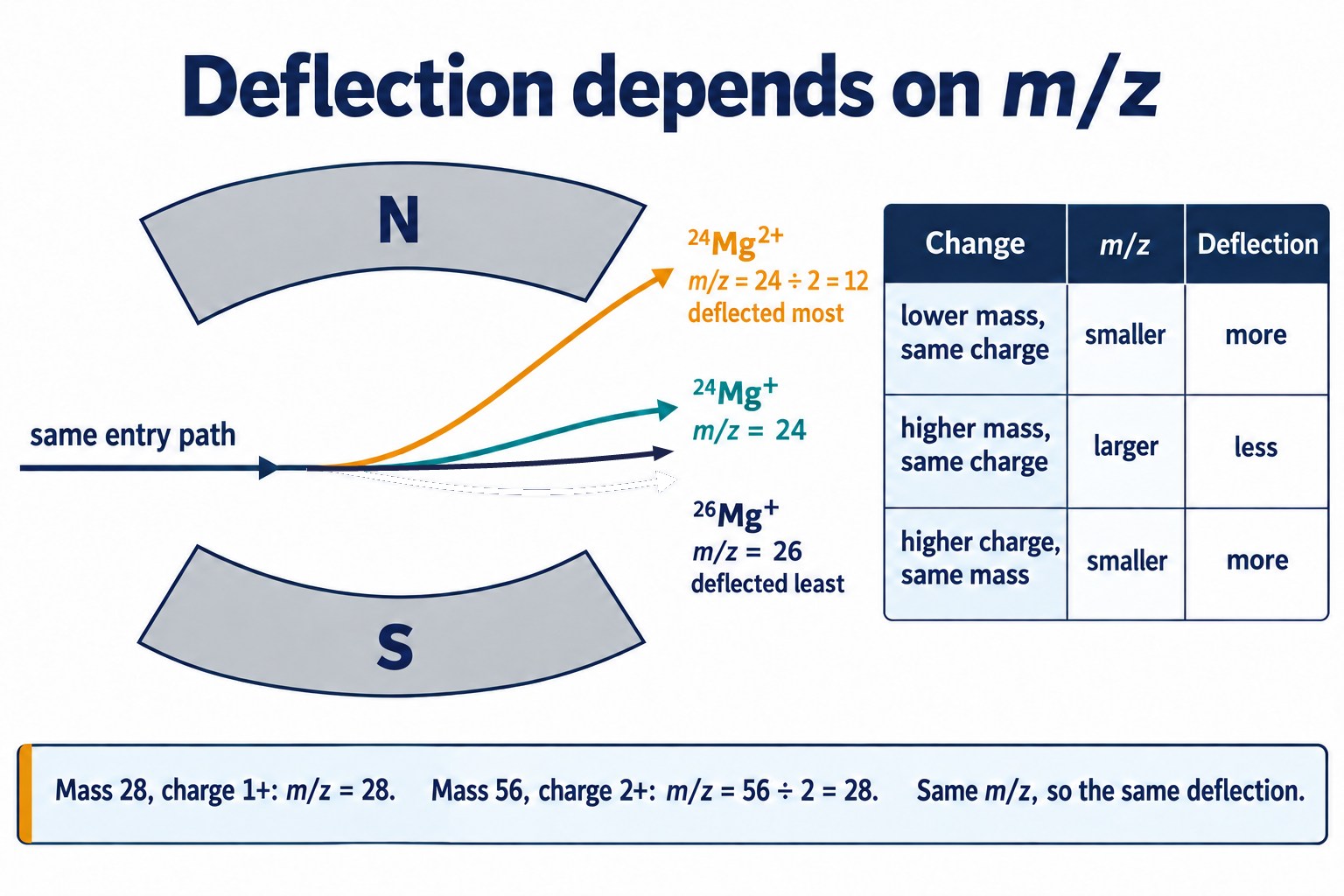
Smaller m/z means more deflection: a 2+ ion has half the m/z of the 1+ ion of the same mass.
Quick Check: Compare the m/z Values
Compare the m/z values and deflections of ions that are not on this page.
Stage 4: Detection
The deflected ions reach a detector plate. When a positive ion hits the detector, it gains an electron and becomes neutral.
Electrons flow from the detector to the positive ions, creating a small electrical current. This signal is amplified and sent to a computer.
The size of the detector signal is proportional to the number of ions reaching the detector. This is why the peak height or intensity in a mass spectrum shows relative abundance.
Exam focus: More ions hitting the detector produce a larger current, so the signal represents greater abundance.
Quick Check: Reading the Detector Current
Use the detector currents to compare two isotopes.
Changing the Magnetic Field
For a fixed magnetic field strength, only ions with a particular m/z value follow the correct path through the slit and reach the detector.
The magnetic field strength is then changed so that ions with different m/z values can be detected.
Ions with a larger m/z are deflected less, so a stronger magnetic field is needed to bend them onto the detector. A weaker field brings ions with a smaller m/z onto the detector.
The computer combines these detector signals to produce the mass spectrum.
Key idea: Changing the magnetic field lets the instrument scan across different m/z values.
Quick Check: Scanning to a Heavier Ion
Decide how the magnetic field must change to detect a heavier ion.
Quick Check: Mass Spectrometer Round-Up
Pick the one fully correct statement in each round to pull the whole page together.
Common Exam Points
Mass spectrometry questions often test precise sequencing, the role of the vacuum, the meaning of m/z, and the link between detector current and abundance.
Use the correct sequence
Vaporise the sample if needed, ionise it, accelerate the positive ions, deflect them in a magnetic field, then detect them.
State why a vacuum is needed
A vacuum prevents collisions with air particles and prevents air particles from becoming ionised and detected.
Do not use mass alone when charge can vary
Deflection depends on m/z, not only mass. A higher charge lowers the m/z value.
Link current to abundance
A larger current is produced when more ions reach the detector, so signal intensity is proportional to abundance.
QuickSnap
In a mass spectrometer, the sample is first vaporised if needed, then ionised by electron bombardment to form positive ions.
The positive ions are accelerated by an electric field and deflected by a magnetic field according to their m/z value. Ions with smaller m/z values are deflected more, while ions with larger m/z values are deflected less.
When ions reach the detector, they gain electrons and produce a current. The size of this current is proportional to the abundance of the ions reaching the detector.
Ionisation
High-energy electrons remove electrons from gaseous particles, forming positive ions.
Acceleration
Positive ions are attracted towards a negative electric field and form a beam.
Deflection
The magnetic field separates ions by m/z. Smaller m/z means greater deflection.
Detection
Positive ions gain electrons at the detector, producing a current proportional to abundance.
The Mass Spectrometer FAQs
Use these quick answers to secure the key wording for Edexcel A Level Chemistry.
What does a mass spectrometer measure?
It separates and detects positive ions according to their mass-to-charge ratio, m/z.
Why must the sample be vaporised?
The sample must be in the gaseous state so that its particles can move through the instrument and be ionised.
How are positive ions formed in electron bombardment?
High-energy electrons collide with gaseous atoms or molecules and remove electrons, producing positive ions.
Why is a vacuum needed in a mass spectrometer?
A vacuum prevents collisions with air particles and prevents air particles from becoming ionised and detected.
What determines the amount of deflection?
The amount of deflection depends on m/z. Ions with smaller m/z values are deflected more.
What does the detector signal show?
The detector signal is a small current. Its size is proportional to the abundance of ions reaching the detector.

Copyright notice: This OLS revision content, including the explanations, layout, diagrams, tables and embedded learning structure, is authored for Online Learning System by Dr. Mohammed Al-Fatah. It may not be copied, reproduced, redistributed or adapted without written permission.
Keep this note — free
Save your progress across every Edexcel topic. A free account remembers which topics you have covered, saves your question scores, and syncs across your phone and laptop.
- Track every topic you have finished
- Keep your practice-question scores
- No payment, no card, free forever
Already registered? Log in